New genes
born by accident
lead to evolutionary innovation
Evolution, Genetics
Novel genes are continuously emerging during evolution, but what drives this process?
A new study, published in PLOS Genetics, has found that the fortuitous appearance of certain combinations of elements in the genome can lead to the generation of new genes.
This work was led by Jorge Ruiz-Orera and Mar Alba from Hospital del Mar Medical Research Institute in Barcelona (IMIM-ICREA).
A new study has found that the fortuitous appearance of certain combinations of elements in the genome can lead to the generation of new genes [Credit: National Human Genome Research Institute]
In every genome, there are sets of genes, which are unique to that particular species.
In this study, the scientists first identified thousands of genes that were specific to human or chimpanzee.
Then, they searched the macaque genome and discovered that this species had significantly less element motifs in the corresponding genomic sequences.
These motifs are recognized by proteins that activate gene expression, a necessary step in the formation of a new gene. The formation of genes de novo from previously non-active parts of the genome was, until recently, considered highly improbable.
This study has shown that the mutations that occur normally in our genetic material may be sufficient to explain how this happens.
Once expressed, the genes can act as a substrate for the evolution of new molecular functions.
This study identified several candidate human proteins that bear no resemblance to any other known protein.
What they do is an enigma still to be resolved.
Source: PLOS [December 31, 2015]
Visualizzazione post con etichetta Evolution. Mostra tutti i post
Visualizzazione post con etichetta Evolution. Mostra tutti i post
martedì 27 ottobre 2015
Geni Mobili ed Evoluzione.
Scientists discover protein factories
hidden in
human jumping genes
Breakingnews, Evolution, Genetics, Human Evolution
Scientists have discovered a previously unknown wellspring of genetic diversity in humans, chimps and most other primates.
This diversity arises from a new component of itinerant sections of genetic code known as jumping genes.
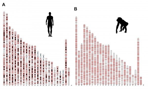
In a paper published October 22, 2015 in Cell, Salk scientists report finding human and chimp DNA peppered with sequences of genetic code they've dubbed ORF0, which spreads throughout the genome on jumping genes.
The ORF0 sequences may produce hundreds or even thousands of previously unknown proteins.
The abundance of ORF0 instances in the human genome suggests that it played--and still plays--an important role in evolutionary diversity and flexibility by serving as a mechanism for generating novel proteins. The discovery of these mobile protein factories may also shine light on the origins of genetic mutations responsible for cancer, mental disorders and other diseases.
"This discovery shows that jumping genes are an even more important source of variation in the primate genome than we thought, whether you're looking at the level of different species, different people or even the different cells within an individual's body," says Rusty Gage, senior author of the paper and a professor in Salk's Laboratory of Genetics.
With the sequencing of the human genome, it became clear that jumping genes--mobile genetic elements first discovered in maize by Barbara McClintock in the early 1950s--were also present and highly active during human evolution.
About half of the human genome resulted from sequences of genetic code that moved or insert extra copies of themselves throughout the genome.
The evolutionary importance of jumping genes was highlighted by the results of another recent study by Gage and collaborators at Stanford. The research used stem cell technologies developed in Gage's lab to explore how differences in gene expression contribute to human and chimp facial structure.
The findings, also reported in Cell, suggested that jumping genes played a role in the evolutionary split between humans and other primates.
In the more recent study that uncovered ORF0, Gage and his colleagues focused on a class of jumping genes known as LINE-1 elements, which make up about 17 percent of the human genome.
These elements contain all the necessary genetic machinery for moving themselves and other classes of jumping genes, unaided, to elsewhere in the genome.
Previously, it was thought that LINE-1 elements contain just two sequences that coded for proteins, the ultimate product of genes that serve a wide range of roles in our cells and organs.
These sequences are known as open reading frames (ORF), and the two previously known sequences, ORF1 and ORF2, are thought to be involved in producing proteins that are important for allowing LINE-1 elements to move around in the genome. In their new study, Gage and his colleagues discovered a third open reading frame. They named it ORF0 based on its location in the LINE-1 element next to ORF1. The scientists found ORF0 in about 3,500 locations in the DNA of humans and about 3,000 locations in the chimp and most other primate genomes.
The structure of LINE-1 elements is such that when an element moves to another site there is a possibility that the ORF0 sequence can blend with genetic sequences in new location in DNA. The result of the new gene sequences can be a new protein. Evolutionarily speaking, this represents a way to generate entirely new molecules that could be beneficial to a species. On the other hand, the reshuffling of an existing ORF0 sequence during a jump could result in a disease-causing mutation.
"This discovery redraws the blueprint of an important piece of genetic machinery in primates, adding a completely new gear," says Ahmet Denli, a staff scientist in Gage's lab and the first author on the paper reporting the findings.
"Jumping genes with ORF0 are basically protein factories with wheels, and over the eons evolution has been driving the bus."
Now that they have identified ORF0 in the primate genome, Denli says they plan to determine how many of the instances of ORF0 actually code for proteins and to investigate what function those proteins serve. The researchers also plan to investigate the behavior of ORF0 in different cell types and diseases. They are particularly interested in exploring its role in cancers and in neurological disorders such as schizophrenia, where previous studies have suggested jumping genes may be involved.
Source: Salk Institute [October 22,2015]

hidden in
human jumping genes
Breakingnews, Evolution, Genetics, Human Evolution
Scientists have discovered a previously unknown wellspring of genetic diversity in humans, chimps and most other primates.
This diversity arises from a new component of itinerant sections of genetic code known as jumping genes.
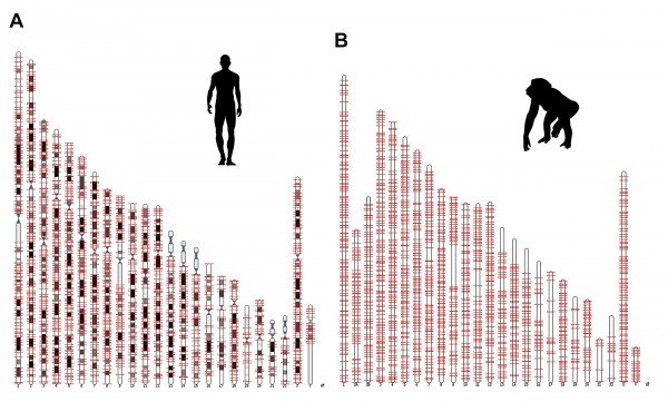
Salk researchers discovered a new genetic component, called ORF0, spread throughout the DNA of humans, chimps and most other primates. This image shows the locations of the ORFO on human and chimp chromosomes
[Credit: Salk Institute]
[Credit: Salk Institute]
In a paper published October 22, 2015 in Cell, Salk scientists report finding human and chimp DNA peppered with sequences of genetic code they've dubbed ORF0, which spreads throughout the genome on jumping genes.
The ORF0 sequences may produce hundreds or even thousands of previously unknown proteins.
The abundance of ORF0 instances in the human genome suggests that it played--and still plays--an important role in evolutionary diversity and flexibility by serving as a mechanism for generating novel proteins. The discovery of these mobile protein factories may also shine light on the origins of genetic mutations responsible for cancer, mental disorders and other diseases.
"This discovery shows that jumping genes are an even more important source of variation in the primate genome than we thought, whether you're looking at the level of different species, different people or even the different cells within an individual's body," says Rusty Gage, senior author of the paper and a professor in Salk's Laboratory of Genetics.
With the sequencing of the human genome, it became clear that jumping genes--mobile genetic elements first discovered in maize by Barbara McClintock in the early 1950s--were also present and highly active during human evolution.
About half of the human genome resulted from sequences of genetic code that moved or insert extra copies of themselves throughout the genome.
The evolutionary importance of jumping genes was highlighted by the results of another recent study by Gage and collaborators at Stanford. The research used stem cell technologies developed in Gage's lab to explore how differences in gene expression contribute to human and chimp facial structure.
The findings, also reported in Cell, suggested that jumping genes played a role in the evolutionary split between humans and other primates.
In the more recent study that uncovered ORF0, Gage and his colleagues focused on a class of jumping genes known as LINE-1 elements, which make up about 17 percent of the human genome.
These elements contain all the necessary genetic machinery for moving themselves and other classes of jumping genes, unaided, to elsewhere in the genome.
Previously, it was thought that LINE-1 elements contain just two sequences that coded for proteins, the ultimate product of genes that serve a wide range of roles in our cells and organs.
These sequences are known as open reading frames (ORF), and the two previously known sequences, ORF1 and ORF2, are thought to be involved in producing proteins that are important for allowing LINE-1 elements to move around in the genome. In their new study, Gage and his colleagues discovered a third open reading frame. They named it ORF0 based on its location in the LINE-1 element next to ORF1. The scientists found ORF0 in about 3,500 locations in the DNA of humans and about 3,000 locations in the chimp and most other primate genomes.
The structure of LINE-1 elements is such that when an element moves to another site there is a possibility that the ORF0 sequence can blend with genetic sequences in new location in DNA. The result of the new gene sequences can be a new protein. Evolutionarily speaking, this represents a way to generate entirely new molecules that could be beneficial to a species. On the other hand, the reshuffling of an existing ORF0 sequence during a jump could result in a disease-causing mutation.
"This discovery redraws the blueprint of an important piece of genetic machinery in primates, adding a completely new gear," says Ahmet Denli, a staff scientist in Gage's lab and the first author on the paper reporting the findings.
"Jumping genes with ORF0 are basically protein factories with wheels, and over the eons evolution has been driving the bus."
Now that they have identified ORF0 in the primate genome, Denli says they plan to determine how many of the instances of ORF0 actually code for proteins and to investigate what function those proteins serve. The researchers also plan to investigate the behavior of ORF0 in different cell types and diseases. They are particularly interested in exploring its role in cancers and in neurological disorders such as schizophrenia, where previous studies have suggested jumping genes may be involved.
Source: Salk Institute [October 22,2015]


sabato 24 ottobre 2015
Biounità costruttiva: la spirale del DNA è sempre la stessa
Tiny DNA building block
is identical
regardless of species
Breakingnews, Evolution, Genetics
The diameter of a single, cylindrical DNA component is always the same, regardless of whether it forms part of a fruit fly, a beech tree, or a human being. This tiny building block in the DNA architecture is part of what is known as the nucleosome. The diameter of the nucleosome has long been a source of fascination for Professor Jakob Bohr of DTU Nanotech at Technical University of Denmark. Because if it is the same in all species, what universal principle or law of nature is in effect?

"As a physicist, I attempt to find the answers to these questions in the worlds of mathematics and physics. And in these contexts, our calculations demonstrate that the size of the nucleosomes makes perfect sense. They create what we have chosen to call 'twist neutrality'," relates Jakob Bohr, who has been studying the diameter of the nucleosomes in partnership with his colleague, Kasper Olsen. "Twist neutrality ensures that the number of turns in the DNA material always remains constant, such that the smallest structure in DNA architecture -- the DNA helix -- can be stretched without affecting the number of twists. This property is essential in nature every time DNA material needs to be copied; in the case of cell division, for example. Briefly put, twist neutrality helps prevent the twists becoming entangled and damaging our DNA," he adds. The nucleosome is only a tiny part of the complex DNA architecture. In total, every single sell contains more than a metre of DNA, and to make room for so much material, the DNA is twisted and folded together over and over again. This leads to the creation of what can be perceived as different levels of structuring, and depending on how a level can actually be defined, eight such levels are created. "Each level alters the scale of our hereditary material by a factor of around three, in the same way as when you fold a piece of paper repeatedly, the thickness is increased by a factor of two every time. This compression of the DNA ensures that it is small enough to fit inside the nucleus of the cell," explains Jakob Bohr. The highest level of DNA comprises the chromosome structures, where the gender chromosomes -- X and Y -- are the best known. The bottom level, i.e. the tiniest structure in the DNA architecture, is the famous double helix, the eye-catching spiral of two strands of DNA winding around one another. The natural law of the twists At the level before this, the double helix is wound twice around a collection of proteins. This is the structure that is called a nucleosome. It takes a lot of nucleosomes to wind up the entire double helix, and this makes the structure resemble a string of pearls -- where the nucleosomes are the pearls and the double helix is the chain. The calculations performed by Jakob Bohr and his colleagues demonstrate that if the diameter of the nucleosomes were different, twist neutrality would simply not exist. "This would not be good, because it would mean that new, inappropriate structures could appear in the DNA. So we think that the reason why the size of the nucleosomes remains constant across species is that there is no benefit to nature in developing or changing this size. Otherwise, we would probably have encountered it as a stage in the evolutionary process," explains Jakob Bohr. New technology should help The long-term aim of the research into the diameter of the nucleosomes is to contribute to a complete description of the geometry of DNA. This could help reveal general principles for the construction of the chromosomes, which would enable us to understand them in even greater detail. "Because even though we can already mine down to gene level in the double helix -- the lowest level in the chromosome -- a number of the other eight levels are still largely uncharted territory. Simply put, we have no idea what structures exist on some of the other levels," adds Jakob Bohr. According to Jakob Bohr, researchers do not currently have access to technology with the capacity to clarify the structures completely at all levels of the chromosomes. However, he is sure that the technology required should appear over the coming 10-15 years, possibly as a result of other research into bioinformatics and via the new facilities including ESS and MAX IV in Lund. Author: Lotte Krull | Source: Technical University of Denmark [October 21, 2015]
is identical
regardless of species
Breakingnews, Evolution, Genetics
The diameter of a single, cylindrical DNA component is always the same, regardless of whether it forms part of a fruit fly, a beech tree, or a human being. This tiny building block in the DNA architecture is part of what is known as the nucleosome. The diameter of the nucleosome has long been a source of fascination for Professor Jakob Bohr of DTU Nanotech at Technical University of Denmark. Because if it is the same in all species, what universal principle or law of nature is in effect?

The illustration shows a very simplified, comic strip view of the organization of DNA. The organization serves to compress the over one-metre long DNA, so it can fit inside the cell nucleus. If you ‘read’ the figure backwards—i.e. from H to A—it corresponds to looking at a chromosome, then ‘zooming in’' step-by-step, so you finally end with the chromosome’s smallest structure—the DNA helix in Figure A.
Figure A The well-known double helix—the spiral with the two strands of DNA is the smallest structure in the chromosome. Here it is reproduced without the base pairs that connect the two strands and which cause the spiral to resemble a spiral ladder.
Figure B shows how the double helix is wound twice around a cylindrical core of proteins. This structure is called a nucleosome. It is the diameter of this structure which is the same across different species—and which fascinates Professor Jakob Bohr. The proportions in this figure do not correspond to the real world, where the DNA helix is equal in size to the protein core.
Figure C shows the row of nucleosomes, causing the structure to resemble a chain of pearls. Figure D-G illustrates the next levels, where the material DNA is further packaged. As yet, the geometry of these levels is not fully clarified.
Level H illustrates the contours of the chromosome as viewed under a microscope
[Credit: Technical University of Denmark]
Level H illustrates the contours of the chromosome as viewed under a microscope
[Credit: Technical University of Denmark]
"As a physicist, I attempt to find the answers to these questions in the worlds of mathematics and physics. And in these contexts, our calculations demonstrate that the size of the nucleosomes makes perfect sense. They create what we have chosen to call 'twist neutrality'," relates Jakob Bohr, who has been studying the diameter of the nucleosomes in partnership with his colleague, Kasper Olsen. "Twist neutrality ensures that the number of turns in the DNA material always remains constant, such that the smallest structure in DNA architecture -- the DNA helix -- can be stretched without affecting the number of twists. This property is essential in nature every time DNA material needs to be copied; in the case of cell division, for example. Briefly put, twist neutrality helps prevent the twists becoming entangled and damaging our DNA," he adds. The nucleosome is only a tiny part of the complex DNA architecture. In total, every single sell contains more than a metre of DNA, and to make room for so much material, the DNA is twisted and folded together over and over again. This leads to the creation of what can be perceived as different levels of structuring, and depending on how a level can actually be defined, eight such levels are created. "Each level alters the scale of our hereditary material by a factor of around three, in the same way as when you fold a piece of paper repeatedly, the thickness is increased by a factor of two every time. This compression of the DNA ensures that it is small enough to fit inside the nucleus of the cell," explains Jakob Bohr. The highest level of DNA comprises the chromosome structures, where the gender chromosomes -- X and Y -- are the best known. The bottom level, i.e. the tiniest structure in the DNA architecture, is the famous double helix, the eye-catching spiral of two strands of DNA winding around one another. The natural law of the twists At the level before this, the double helix is wound twice around a collection of proteins. This is the structure that is called a nucleosome. It takes a lot of nucleosomes to wind up the entire double helix, and this makes the structure resemble a string of pearls -- where the nucleosomes are the pearls and the double helix is the chain. The calculations performed by Jakob Bohr and his colleagues demonstrate that if the diameter of the nucleosomes were different, twist neutrality would simply not exist. "This would not be good, because it would mean that new, inappropriate structures could appear in the DNA. So we think that the reason why the size of the nucleosomes remains constant across species is that there is no benefit to nature in developing or changing this size. Otherwise, we would probably have encountered it as a stage in the evolutionary process," explains Jakob Bohr. New technology should help The long-term aim of the research into the diameter of the nucleosomes is to contribute to a complete description of the geometry of DNA. This could help reveal general principles for the construction of the chromosomes, which would enable us to understand them in even greater detail. "Because even though we can already mine down to gene level in the double helix -- the lowest level in the chromosome -- a number of the other eight levels are still largely uncharted territory. Simply put, we have no idea what structures exist on some of the other levels," adds Jakob Bohr. According to Jakob Bohr, researchers do not currently have access to technology with the capacity to clarify the structures completely at all levels of the chromosomes. However, he is sure that the technology required should appear over the coming 10-15 years, possibly as a result of other research into bioinformatics and via the new facilities including ESS and MAX IV in Lund. Author: Lotte Krull | Source: Technical University of Denmark [October 21, 2015]
domenica 27 settembre 2015
Nuova Sintesi
New synthesis method
imitates the way molecules were formed
at the dawn of life on Earth

This is a significant step forward since it replicates the formation of carbohydrates in conditions resembling those that presumably initiated life on Earth (prebiotic conditions) and because it allows relatively large organic molecules to be obtained very selectively and efficiently.
Furthermore, it is a process with few steps, that does not use organic solvents and generates no waste, and it has great potential in chemistry, especially for obtaining molecules and active ingredients of interest (drugs, supplements, etc.).
Pere Clapés, a research professor with the CSIC who led this project, explains that in the synthesis of organic molecules . "it is not only important for them to have the correct structure, but also the right angle and position in space, because this affects their function."
In fact, this is one of the main problems that can limit the effectiveness of compounds like drugs.
In the case of pentoses and hexoses, these are simple sugars (monosaccharides) with five and six carbon atoms, respectively: crucial for life thanks to their function in energy production, structuring, communication and cell-cell recognition.
The results presented in the journal Nature Chemistry show that the scientists obtained pentoses and hexoses by assembling formaldehyde and glycolaldehyde, with a minimal modification to the FSA enzyme sequence.
A very malleable enzyme
The enzyme FSA was discovered in 2001 and its physiological function in E. coli is still unknown.
It is thought to be an ancestral enzyme, and that it is active before a broad range of compounds.
What surprised the researchers is that it is a very malleable enzyme, much more so than others.
As a result, with only a small number of genetic mutations in the enzyme, its catalytic capacity can be modulated and increased significantly.
This is what allows the enzyme to be carefully adapted in order to synthesise several molecules at will.
The metabolism of carbohydrates in living organisms is a complex process, forged over millions of years of evolution.
It is no easy task to carry out these processes in a flask, whether by assembling the enzymes involved in the process or by manipulating the metabolic pathways of living organisms.
Nor is it simple to obtain carbohydrates with conventional chemical methods, which require several stages and the use of organic solvents. The procedure was developed by scientists in the Biotransformation and Active Molecules Group of the Spanish National Research Council (CSIC), with support from the Nuclear Magnetic Resonance Service of the UAB.
Pere Clapés explains: "we want to prove that the tools of biocatalysis allow complex molecules to be obtained from simpler ones, which are in fact the same ones used in nature."
He goes on: "Over millions of years, living organisms have forged these metabolic strategies to obtain the carbohydrates they need to survive." "The process is a simple one, mimicking the prebiotic formation of carbohydrates from compounds that were probably around in the world before life began," adds Teodor Parella, of the UAB.
For these researchers, the engineering of proteins, in particular of biocatalysts, has enormous potential for the sustainable synthesis of natural molecules and their derived products.
Source: Universitat Autònoma de Barcelona [September 23, 2015]
imitates the way molecules were formed
at the dawn of life on Earth
Breakingnews, Evolution, Genetics, Origin of Life

Researchers from the Institute for Advanced Chemistry of Catalonia (IQAC-CSIC), with support from the Nuclear Magnetic Resonance Service of the Universitat Autònoma de Barcelona (UAB) have developed a method for synthesising organic molecules very selectively, by assembling simple molecules and using an enzyme from E. coli (FSA: D-
fructose-6-phosphate aldolase), which acts as a biocatalyst. E-coli enzyme
[Credit: CSIC]
This is a significant step forward since it replicates the formation of carbohydrates in conditions resembling those that presumably initiated life on Earth (prebiotic conditions) and because it allows relatively large organic molecules to be obtained very selectively and efficiently.
Furthermore, it is a process with few steps, that does not use organic solvents and generates no waste, and it has great potential in chemistry, especially for obtaining molecules and active ingredients of interest (drugs, supplements, etc.).
Pere Clapés, a research professor with the CSIC who led this project, explains that in the synthesis of organic molecules . "it is not only important for them to have the correct structure, but also the right angle and position in space, because this affects their function."
In fact, this is one of the main problems that can limit the effectiveness of compounds like drugs.
In the case of pentoses and hexoses, these are simple sugars (monosaccharides) with five and six carbon atoms, respectively: crucial for life thanks to their function in energy production, structuring, communication and cell-cell recognition.
The results presented in the journal Nature Chemistry show that the scientists obtained pentoses and hexoses by assembling formaldehyde and glycolaldehyde, with a minimal modification to the FSA enzyme sequence.
A very malleable enzyme
The enzyme FSA was discovered in 2001 and its physiological function in E. coli is still unknown.
It is thought to be an ancestral enzyme, and that it is active before a broad range of compounds.
What surprised the researchers is that it is a very malleable enzyme, much more so than others.
As a result, with only a small number of genetic mutations in the enzyme, its catalytic capacity can be modulated and increased significantly.
This is what allows the enzyme to be carefully adapted in order to synthesise several molecules at will.
The metabolism of carbohydrates in living organisms is a complex process, forged over millions of years of evolution.
It is no easy task to carry out these processes in a flask, whether by assembling the enzymes involved in the process or by manipulating the metabolic pathways of living organisms.
Nor is it simple to obtain carbohydrates with conventional chemical methods, which require several stages and the use of organic solvents. The procedure was developed by scientists in the Biotransformation and Active Molecules Group of the Spanish National Research Council (CSIC), with support from the Nuclear Magnetic Resonance Service of the UAB.
Pere Clapés explains: "we want to prove that the tools of biocatalysis allow complex molecules to be obtained from simpler ones, which are in fact the same ones used in nature."
He goes on: "Over millions of years, living organisms have forged these metabolic strategies to obtain the carbohydrates they need to survive." "The process is a simple one, mimicking the prebiotic formation of carbohydrates from compounds that were probably around in the world before life began," adds Teodor Parella, of the UAB.
For these researchers, the engineering of proteins, in particular of biocatalysts, has enormous potential for the sustainable synthesis of natural molecules and their derived products.
Source: Universitat Autònoma de Barcelona [September 23, 2015]
domenica 5 luglio 2015
La Peste e la Storia
How small genetic change in Yersinia pestis changed human history
Breakingnews, Evolution, Genetics
While studying Yersinia pestis, the bacteria responsible for epidemics of plague such as the Black Death, Wyndham Lathem, Ph.D., assistant professor in microbiology-immunology at Northwestern University Feinberg School of Medicine, found a single small genetic change that fundamentally influenced the evolution of the deadly pathogen, and thus the course of human history.


In a paper published in Nature Communications, Lathem and first author Daniel Zimbler, Ph.D., a Feinberg post-doctoral fellow, demonstrated how the acquisition of a single gene caused the shift of Y. pestis from causing a primarily gastrointestinal infection to a more serious and often fatal respiratory disease.
They further showed how later modifications of this gene enhanced infections associated with the bubonic plague.
"Our findings demonstrate how Y. pestis had the ability to cause a severe respiratory disease very early in its evolution. This research helps us better understand how bacteria can adapt to new host environments to cause disease by acquiring small bits of DNA," Lathem said. The team examined ancestral strains of the bacteria in mouse models to learn when Y. pestis gained the ability to infect the lungs and cause the severe form of the disease known as pneumonic plague. In the most ancestral of all currently existing Y. pestis strains, they showed how the bacteria could successfully colonize the lungs but could not cause the severe disease associated with pneumonic plague. The biggest difference they found between this strain and closely related strains that could cause pneumonic plague was a gene for the surface protein Pla.
Lathem proposed that the bacteria's acquisition of the gene Pla enhanced its ability to cause infection in the lungs and was all that this ancestral strain of Y. pestis needed to produce a fatal lung infection.
So Lathem and his team inserted the Pla gene into this strain to observe changes in the health of the lungs. They found the newly mutated strain had gained the ability to cause respiratory infection identically to modern strains of Y. pestis that cause disease today, demonstrating that the Pla gene was necessary for Y. pestis to infect the lungs. In addition, they found that no other changes to Y. pestis were required, even though the bacteria has continued to gain and lose genes over the last several thousand years. The lab also looked at variations of the gene Pla and discovered that a single modification only found in modern strains of Y. pestis was a critical adaptation for the bacteria to spread in the body and infect the lymph nodes, a form of the infection that causes bubonic plague. According to Lathem, the surprising conclusion from this aspect of the study is that, contrary to current thinking in the field, Y. pestis may have first evolved as a respiratory pathogen before it could cause the more common form of disease, bubonic plague. Lathem said the new research may explain how Y. pestis transitioned from causing only localized outbreaks of plague to the pandemic spread of Y. pestis such as the sixth century's Justinian Plague and the fourteenth century's Black Death. "Our data suggests that the insertion and then subsequent mutation of Pla allowed for new, rapidly evolving strains of disease," Lathem said. "This information can show how new respiratory pathogens could emerge with only small genetic changes."
Author: Sarah Plumridge | Source: Northwestern University [June 30, 2015]
Breakingnews, Evolution, Genetics
While studying Yersinia pestis, the bacteria responsible for epidemics of plague such as the Black Death, Wyndham Lathem, Ph.D., assistant professor in microbiology-immunology at Northwestern University Feinberg School of Medicine, found a single small genetic change that fundamentally influenced the evolution of the deadly pathogen, and thus the course of human history.


This image of lung tissue shows inflammatory lesions in an ancestral strain of the bacteria that causes pneumonic plague without the Pla gene (above) and with the gene (below). The addition of Pla allowed this strain to cause pneumonic plague
[Credit: Northwestern University]
[Credit: Northwestern University]
In a paper published in Nature Communications, Lathem and first author Daniel Zimbler, Ph.D., a Feinberg post-doctoral fellow, demonstrated how the acquisition of a single gene caused the shift of Y. pestis from causing a primarily gastrointestinal infection to a more serious and often fatal respiratory disease.
They further showed how later modifications of this gene enhanced infections associated with the bubonic plague.
"Our findings demonstrate how Y. pestis had the ability to cause a severe respiratory disease very early in its evolution. This research helps us better understand how bacteria can adapt to new host environments to cause disease by acquiring small bits of DNA," Lathem said. The team examined ancestral strains of the bacteria in mouse models to learn when Y. pestis gained the ability to infect the lungs and cause the severe form of the disease known as pneumonic plague. In the most ancestral of all currently existing Y. pestis strains, they showed how the bacteria could successfully colonize the lungs but could not cause the severe disease associated with pneumonic plague. The biggest difference they found between this strain and closely related strains that could cause pneumonic plague was a gene for the surface protein Pla.
Lathem proposed that the bacteria's acquisition of the gene Pla enhanced its ability to cause infection in the lungs and was all that this ancestral strain of Y. pestis needed to produce a fatal lung infection.
So Lathem and his team inserted the Pla gene into this strain to observe changes in the health of the lungs. They found the newly mutated strain had gained the ability to cause respiratory infection identically to modern strains of Y. pestis that cause disease today, demonstrating that the Pla gene was necessary for Y. pestis to infect the lungs. In addition, they found that no other changes to Y. pestis were required, even though the bacteria has continued to gain and lose genes over the last several thousand years. The lab also looked at variations of the gene Pla and discovered that a single modification only found in modern strains of Y. pestis was a critical adaptation for the bacteria to spread in the body and infect the lymph nodes, a form of the infection that causes bubonic plague. According to Lathem, the surprising conclusion from this aspect of the study is that, contrary to current thinking in the field, Y. pestis may have first evolved as a respiratory pathogen before it could cause the more common form of disease, bubonic plague. Lathem said the new research may explain how Y. pestis transitioned from causing only localized outbreaks of plague to the pandemic spread of Y. pestis such as the sixth century's Justinian Plague and the fourteenth century's Black Death. "Our data suggests that the insertion and then subsequent mutation of Pla allowed for new, rapidly evolving strains of disease," Lathem said. "This information can show how new respiratory pathogens could emerge with only small genetic changes."
Author: Sarah Plumridge | Source: Northwestern University [June 30, 2015]
giovedì 28 maggio 2015
Dal Pollo al Dinosauro
Sono noti pochissimi esperimenti che riportano la morfologia e la meccanica degli attuali uccelli indietro , fino alle modalità che erano proprie dei dinosauri.
Ecco perché quello riportato qui è un esempio prezioso per gli scienziati. I dinosauri avevano zampe con un "alluce" non opponibile, sito indietro ed in alto (come appare essere nei cani e nei gatti oggi). Negli uccelli, invece, si trova più in basso (perché scende durante lo sviluppo embrionale, grazie all'attività muscolare locale embrionale) e permette loro di 'appollaiarsi', con l'aggrapparsi intorno ad un ramo, oppure di 'ghermire' la preda con un solo gesto.
Bloccando tali muscoli nell'embrione, si ottengono polli con zampe 'da dinosauro'. Un altro esperimento che dimostra attraverso quali complicate strade anche biologiche debba procedere l'informazione genetica per potere avere il proprio effetto finale.
From chicken to dinosaur:
Scientists experimentally reverse the evolution of the perching toe
A unique adaptation in the foot of birds is the presence of a thumb-like opposable toe, which allows them to grasp and perch.
However, in their dinosaur ancestors, this toe was small and non- opposable, and did not even touch the ground, resembling the dewclaws of dogs and cats.

Remarkably, the embryonic development of birds provides a parallel of this evolutionary history: The toe starts out like their dinosaur ancestors, but then its base (the metatarsal) becomes twisted, making it opposable.
Brazilian researcher Joâo Botelho, working at the lab of Alexander Vargas at the University of Chile, decided to study the underlying mechanisms.
Botelho observed that the twisting occurred shortly after the embryonic musculature of this toe was in place.
"This is one of the clearest examples of how indirect the morphological consequences of genetic change are mediated," Gunter Wagner, evolutionary geneticist and professor at Yale.
Bird embryos move a lot inside the egg during development, and the onset of movement at this toe coincided with the twisting of its base.
Botelho also demonstrated that in this toe, genes of cartilage maturation were expressed at a much later stage than other digits: It retains many rapidly dividing stem cells for a much longer period.
Such immature cartilage is highly plastic and easily transformed by muscular activity.
These observations suggested the toe is twisted as a result of mechanical forces imposed on it by the embryonic musculature.
Definitive proof, however, would come from experiments.
When Botelho applied Decamethonium bromide, a pharmacological agent capable of paralyzing embryonic musculature, the result was a non-opposable toe with a straight, non-twisted base identical to that of their dinosaur ancestors.
Only a few experiments are known to recover dinosaur traits in birds (such as a dinosaur-like shank and tooth-like structures).
The undoing of the perching digit is thus an important new addition, and the results have now been published in Scientific Reports.
The significance of this experiment, however, goes beyond the fact that a dinosaur-like toe is being retrieved.
Evolutionary research often centers on mutations, but the development and evolution of the perching toe cannot be understood without the forces of embryonic muscular activity.
The study is described as "true developmental mechanics" by Gunter Wagner, an evolutionary geneticist and professor at Yale.
"This is one of the clearest examples of how indirect the morphological consequences of genetic change are mediated.
The experiments prove that interactions about organ systems channel the directions of organismal evolution."
Source: Universidad de Chile [May 22, 2015]
Ecco perché quello riportato qui è un esempio prezioso per gli scienziati. I dinosauri avevano zampe con un "alluce" non opponibile, sito indietro ed in alto (come appare essere nei cani e nei gatti oggi). Negli uccelli, invece, si trova più in basso (perché scende durante lo sviluppo embrionale, grazie all'attività muscolare locale embrionale) e permette loro di 'appollaiarsi', con l'aggrapparsi intorno ad un ramo, oppure di 'ghermire' la preda con un solo gesto.
Bloccando tali muscoli nell'embrione, si ottengono polli con zampe 'da dinosauro'. Un altro esperimento che dimostra attraverso quali complicate strade anche biologiche debba procedere l'informazione genetica per potere avere il proprio effetto finale.
From chicken to dinosaur:
Scientists experimentally reverse the evolution of the perching toe
A unique adaptation in the foot of birds is the presence of a thumb-like opposable toe, which allows them to grasp and perch.
However, in their dinosaur ancestors, this toe was small and non- opposable, and did not even touch the ground, resembling the dewclaws of dogs and cats.

The perching toe from chicken to dinosaur
[Credit: Universidad de Chile]
Remarkably, the embryonic development of birds provides a parallel of this evolutionary history: The toe starts out like their dinosaur ancestors, but then its base (the metatarsal) becomes twisted, making it opposable.
Brazilian researcher Joâo Botelho, working at the lab of Alexander Vargas at the University of Chile, decided to study the underlying mechanisms.
Botelho observed that the twisting occurred shortly after the embryonic musculature of this toe was in place.
"This is one of the clearest examples of how indirect the morphological consequences of genetic change are mediated," Gunter Wagner, evolutionary geneticist and professor at Yale.
Bird embryos move a lot inside the egg during development, and the onset of movement at this toe coincided with the twisting of its base.
Botelho also demonstrated that in this toe, genes of cartilage maturation were expressed at a much later stage than other digits: It retains many rapidly dividing stem cells for a much longer period.
Such immature cartilage is highly plastic and easily transformed by muscular activity.
These observations suggested the toe is twisted as a result of mechanical forces imposed on it by the embryonic musculature.
Definitive proof, however, would come from experiments.
When Botelho applied Decamethonium bromide, a pharmacological agent capable of paralyzing embryonic musculature, the result was a non-opposable toe with a straight, non-twisted base identical to that of their dinosaur ancestors.
Only a few experiments are known to recover dinosaur traits in birds (such as a dinosaur-like shank and tooth-like structures).
The undoing of the perching digit is thus an important new addition, and the results have now been published in Scientific Reports.
The significance of this experiment, however, goes beyond the fact that a dinosaur-like toe is being retrieved.
Evolutionary research often centers on mutations, but the development and evolution of the perching toe cannot be understood without the forces of embryonic muscular activity.
The study is described as "true developmental mechanics" by Gunter Wagner, an evolutionary geneticist and professor at Yale.
"This is one of the clearest examples of how indirect the morphological consequences of genetic change are mediated.
The experiments prove that interactions about organ systems channel the directions of organismal evolution."
Source: Universidad de Chile [May 22, 2015]
lunedì 25 maggio 2015
Il Primo Amico dell'Uomo
Our bond with dogs
may go back more than 27,000 years
Dogs' special relationship to humans may go back 27,000 to 40,000 years, according to genomic analysis of an ancient Taimyr wolf bone reported in the Cell Press journal Current Biology on May 21.
Earlier genome-based estimates have suggested that the ancestors of modern-day dogs diverged from wolves no more than 16,000 years ago, after the last Ice Age.

Ancient Taimyr Wolf bone from the lower jaw. The animal lived approximately 27,000 to 40,000 years ago
[Credit: Love Dalén]
The genome from this ancient specimen, which has been radiocarbon dated to 35,000 years ago, reveals that the Taimyr wolf represents the most recent common ancestor of modern wolves and dogs.
"Dogs may have been domesticated much earlier than is generally believed," says Love Dalén of the Swedish Museum of Natural History.
"The only other explanation is that there was a major divergence between two wolf populations at that time, and one of these populations subsequently gave rise to all modern wolves."
Dalén considers this second explanation less likely, since it would require that the second wolf population subsequently became extinct in the wild.
"It is [still] possible that a population of wolves remained relatively untamed but tracked human groups to a large degree, for a long time," adds first author of the study Pontus Skoglund of Harvard Medical School and the Broad Institute.

DNA from this small piece of a rib bone from an ancient Taimyr wolf suggests that dogs may have been domesticated 27,000 years ago
[Credit: Love Dalén]
The researchers made these discoveries based on a small piece of bone picked up during an expedition to the Taimyr Peninsula in Siberia.
Initially, they didn't realize the bone fragment came from a wolf at all; this was only determined using a genetic test back in the laboratory.
But wolves are common on the Taimyr Peninsula, and the bone could have easily belonged to a modern-day wolf.
On a hunch, the researchers decided to radiocarbon date the bone anyway.
It was only then that they realized what they had: a 35,000-year-old bone from an ancient Taimyr wolf.
The DNA evidence also shows that modern-day Siberian Huskies and Greenland sled dogs share an unusually large number of genes with the ancient Taimyr wolf.
"The power of DNA can provide direct evidence that a Siberian Husky you see walking down the street shares ancestry with a wolf that roamed Northern Siberia 35,000 years ago," Skoglund says.
To put that in perspective, "this wolf lived just a few thousand years after Neandertals disappeared from Europe and modern humans started populating Europe and Asia."
Source: Cell Press [May 21, 2015]
giovedì 14 maggio 2015
6 - Una sesta base?
L'interrogativo è d'obbligo: sarebbe una scoperta epocale.
Classicamente, le basi sono quattro (adenina, citosina, guanina e timina).
Altre due basi derivate da queste sono la metil citosina e la metil adenina, che sono variazioni delle prime due.
Quando si scoprirono le attività biomolecolari importanti delle metil citosina (mC) essa fu aggiunta come quinta base.
Lo studio attuale, di Manuel esteller dell'Università di Barcellona e appena pubblicato sulla rivista Cell, propone che anche la metil Adenina (mA) sia 'promossa' a base; sarebbe quindi la sesta.
Essa è presente in minime quantità in vari batteri (in cui protegge dall'immissione esterna di materiale nucleare) ed in vari vegetali ed animali inferiori (alghe, vermi, mosche) e la sua funzione sarebbe quella di modulare l'attività di vari geni e di influenzare le cellule staminali.
Tutto sta a vedere se si troverà anche nei mammiferi e - in special modo - nell'uomo.
Sixth DNA's base discovered?
DNA (deoxyribonucleic acid) is the main component of our genetic material. It is formed by combining four parts: A, C, G and T (adenine, cytosine, guanine and thymine), called bases of DNA combine in thousands of possible sequences to provide the genetic variability that enables the wealth of aspects and functions of living beings.

Two more bases: the Methyl- cytosine and Methyl-adenine
In the early 80s, to these four "classic" bases of DNA was added a fifth: the methyl-cytosine (mC) derived from cytosine.
And it was in the late 90's when mC was recognized as the main cause of epigenetic mechanisms: it is able to switch genes on or off depending on the physiological needs of each tissue.
In recent years, interest in this fifth DNA base has increased by showing that alterations in the methyl-cytosine contribute to the development of many human diseases, including cancer.
Today, an article published in Cell by Manel Esteller, director of the Epigenetics and Cancer Biology Program of the Bellvitge Biomedical Research Institute (IDIBELL), ICREA researcher and Professor of Genetics at the University of Barcelona, describes the possible existence of a sixth DNA base, the methyl-adenine (mA), which also help determine the epigenome and would therefore be key in the life of the cells.
In bacteria and in complex organisms "It was known for years that bacteria, evolutionarily very distant living organisms of us, had mA in its genome with a protective function against the insertion of genetic material from other organisms.
But it was believed that this was a phenomenon of primitive cells and it was very static" describes Manel Esteller.
"However, this issue of CELL publishes three papers suggesting that more complex cells called eukaryotes such as the human body cells, also present the sixth DNA base.
These studies suggest that algae, worms and flies possess mA and it acts to regulate the expression of certain genes, thus constituting a new epigenetic mark.
This work has been possible thanks to the development of analytical methods with high sensitivity because levels of mA in described genomes are low.
In addition it seems that mA would play a specific role in stem cells and early stages of development, "explains the researcher.
"Now the challenge we face is to confirm this data and find out whether mammals, including humans, we also have this sixth DNA base, and consider what its role is".
Source: IDIBELL-Bellvitge Biomedical Research Institute [May 11, 2015]
Classicamente, le basi sono quattro (adenina, citosina, guanina e timina).
Altre due basi derivate da queste sono la metil citosina e la metil adenina, che sono variazioni delle prime due.
Quando si scoprirono le attività biomolecolari importanti delle metil citosina (mC) essa fu aggiunta come quinta base.
Lo studio attuale, di Manuel esteller dell'Università di Barcellona e appena pubblicato sulla rivista Cell, propone che anche la metil Adenina (mA) sia 'promossa' a base; sarebbe quindi la sesta.
Essa è presente in minime quantità in vari batteri (in cui protegge dall'immissione esterna di materiale nucleare) ed in vari vegetali ed animali inferiori (alghe, vermi, mosche) e la sua funzione sarebbe quella di modulare l'attività di vari geni e di influenzare le cellule staminali.
Tutto sta a vedere se si troverà anche nei mammiferi e - in special modo - nell'uomo.
Sixth DNA's base discovered?
DNA (deoxyribonucleic acid) is the main component of our genetic material. It is formed by combining four parts: A, C, G and T (adenine, cytosine, guanine and thymine), called bases of DNA combine in thousands of possible sequences to provide the genetic variability that enables the wealth of aspects and functions of living beings.

Methyl-Adenine 3D structure [Credit: IDIBELL]
Two more bases: the Methyl- cytosine and Methyl-adenine
In the early 80s, to these four "classic" bases of DNA was added a fifth: the methyl-cytosine (mC) derived from cytosine.
And it was in the late 90's when mC was recognized as the main cause of epigenetic mechanisms: it is able to switch genes on or off depending on the physiological needs of each tissue.
In recent years, interest in this fifth DNA base has increased by showing that alterations in the methyl-cytosine contribute to the development of many human diseases, including cancer.
Today, an article published in Cell by Manel Esteller, director of the Epigenetics and Cancer Biology Program of the Bellvitge Biomedical Research Institute (IDIBELL), ICREA researcher and Professor of Genetics at the University of Barcelona, describes the possible existence of a sixth DNA base, the methyl-adenine (mA), which also help determine the epigenome and would therefore be key in the life of the cells.
In bacteria and in complex organisms "It was known for years that bacteria, evolutionarily very distant living organisms of us, had mA in its genome with a protective function against the insertion of genetic material from other organisms.
But it was believed that this was a phenomenon of primitive cells and it was very static" describes Manel Esteller.
"However, this issue of CELL publishes three papers suggesting that more complex cells called eukaryotes such as the human body cells, also present the sixth DNA base.
These studies suggest that algae, worms and flies possess mA and it acts to regulate the expression of certain genes, thus constituting a new epigenetic mark.
This work has been possible thanks to the development of analytical methods with high sensitivity because levels of mA in described genomes are low.
In addition it seems that mA would play a specific role in stem cells and early stages of development, "explains the researcher.
"Now the challenge we face is to confirm this data and find out whether mammals, including humans, we also have this sixth DNA base, and consider what its role is".
Source: IDIBELL-Bellvitge Biomedical Research Institute [May 11, 2015]
giovedì 2 aprile 2015
SEQUOIA SEMPERVIRENS, esaploide.
le Sequoie sono tra gli alberi più alti ed inoltre sono organismi viventi tra i più più vecchi nel Mondo. Hanno la capacità - che le distingue dalle altre conifere - di riprodursi dai ceppi tagliati, da tronchi caduti, dalle radici.
Queste straordinarie piante (veri fossili viventi) sono adesso sotto studio per aiutarne la conservazione e la salvaguardia: un tempo, infatti, esse coprivano quasi tutto l'emisfero settentrionale, oggi si reperiscono solamente in alcune valli protette della California e dell'Oregon.
Tra gli studi (condotti da ricercatori della Università della California - Berkeley) vi sono quelli genetici, che hanno permesso di stabilire che la sequoia Sempervirens è esaploide, non diploide (e ciò significa che nel proprio genoma possiede 6 copie dei cromosomi omologhi, non solamente 2).
Ciò determina una maggiore difficoltà dello studio genetico, per via del maggior numero di alleli. se a ciò si aggiunge la possibile presenza di mutazioni, si comprende bene la difficoltà che gli autori incontrano nel ricostruire le linee di discendenza.
Fino ad oggi, su 770 individui, sono stati reperiti 449 cloni differenti. La Sequoia è un organismo arboreo di rapida crescita: questo conferisce una rilevanza economica alla specie, oltre a quella di datazione storica assoluta (dendrocronologia) e botanica.
Questi studi, pertanto, potranno aiutare la gestione di un patrimonio di grande interesse da molti punti di vista...
The shortest DNA sequences reveal insights into the world's tallest trees
Coast redwoods are famous for being the tallest trees in the world, but their height is not the only thing that sets them apart.
Unlike most conifer trees, coast redwoods can reproduce by sprouting from cut stumps, fallen logs, and roots.
Researchers from the University of California, Berkeley, are uncovering important information about patterns of coast redwood clones with a new DNA analysis method that could help forest management and preservation efforts.

The new method, described in a recent issue of Applications of Plant Sciences, will enable scientists to identify clonal lineages and study how clonal diversity varies throughout the geographic range of this species.
Coast redwoods are among the oldest living trees on earth and, although they once occupied much of the northern hemisphere, today's native coast redwood forests can only be found in a 450-mile strip of land along the coast of northern California to southern Oregon.
To date, very little research has been done to elucidate the clonal patterns of coast redwood.
This is partly due to another aspect that sets redwoods apart from other woody plants--they are hexaploid, meaning that there are six copies of each homologous chromosome in the genome, instead of two.
"Genetic analyses of polyploid organisms are difficult due to higher numbers of alleles," explains Lakshmi Narayan, the graduate student who led the UC Berkeley study. "In addition to their hexaploid condition, another challenge in determining the genotypic identity of clonal plants is the possibility of somatic mutation, where a mutation occurs that changes the genotype of an individual in a clonal lineage.
These mutations have the potential to confound genotyping studies seeking to identify the origin of shoots." Narayan designed the new protocol to overcome challenges associated with mutations and with the high genetic copy number in redwood DNA.
Narayan and her colleagues tested the clonal identification protocol by collecting DNA samples from 770 redwoods and successfully identified 449 distinct clones.
A key aspect of the new method is the use of short repeating DNA sequences from different coast redwood tissue types.
The short DNA sequences, known as microsatellites, are present in all living organisms and widely used to distinguish individuals from one another.
The researchers analyzed microsatellite data from coast redwood cambium, leaf, and sprout tissue.
"One novel aspect of our protocol was the use of different tissue types to verify consistent identification of alleles between samples," explains Narayan.
"Requiring that alleles amplified in multiple tissue types from the same individual before scoring them made our protocol more conservative. This potentially reduced potential for misidentification. The other novel aspect of our work was the extensive use of data simulation to test the resolution of our genetic markers and scoring protocol."
Because they are a fast-growing tree species, young redwoods are commercially valued for timber production while ancient redwoods remain protected.
Genetic data produced from the new protocol could help guide sustainable forest management of commercial young-growth forests and also improve efforts to preserve ancient redwood populations.
Says Narayan, "We hope the methods presented in this paper will be applicable to other species, in addition to being useful for genotyping coast redwoods.
Our future research will employ this methodology to study the variation in levels of clonal diversity throughout the species' range, and look at how clonal reproduction may affect the physical structure of redwood forests."
Source: Botanical Society of America [March 27, 2015]
Queste straordinarie piante (veri fossili viventi) sono adesso sotto studio per aiutarne la conservazione e la salvaguardia: un tempo, infatti, esse coprivano quasi tutto l'emisfero settentrionale, oggi si reperiscono solamente in alcune valli protette della California e dell'Oregon.
Tra gli studi (condotti da ricercatori della Università della California - Berkeley) vi sono quelli genetici, che hanno permesso di stabilire che la sequoia Sempervirens è esaploide, non diploide (e ciò significa che nel proprio genoma possiede 6 copie dei cromosomi omologhi, non solamente 2).
Ciò determina una maggiore difficoltà dello studio genetico, per via del maggior numero di alleli. se a ciò si aggiunge la possibile presenza di mutazioni, si comprende bene la difficoltà che gli autori incontrano nel ricostruire le linee di discendenza.
Fino ad oggi, su 770 individui, sono stati reperiti 449 cloni differenti. La Sequoia è un organismo arboreo di rapida crescita: questo conferisce una rilevanza economica alla specie, oltre a quella di datazione storica assoluta (dendrocronologia) e botanica.
Questi studi, pertanto, potranno aiutare la gestione di un patrimonio di grande interesse da molti punti di vista...
The shortest DNA sequences reveal insights into the world's tallest trees
Coast redwoods are famous for being the tallest trees in the world, but their height is not the only thing that sets them apart.
Unlike most conifer trees, coast redwoods can reproduce by sprouting from cut stumps, fallen logs, and roots.
Researchers from the University of California, Berkeley, are uncovering important information about patterns of coast redwood clones with a new DNA analysis method that could help forest management and preservation efforts.
Coast redwoods (Sequioa sempervirens)
[Credit: focus.tracinglight.com]
The new method, described in a recent issue of Applications of Plant Sciences, will enable scientists to identify clonal lineages and study how clonal diversity varies throughout the geographic range of this species.
Coast redwoods are among the oldest living trees on earth and, although they once occupied much of the northern hemisphere, today's native coast redwood forests can only be found in a 450-mile strip of land along the coast of northern California to southern Oregon.
To date, very little research has been done to elucidate the clonal patterns of coast redwood.
This is partly due to another aspect that sets redwoods apart from other woody plants--they are hexaploid, meaning that there are six copies of each homologous chromosome in the genome, instead of two.
"Genetic analyses of polyploid organisms are difficult due to higher numbers of alleles," explains Lakshmi Narayan, the graduate student who led the UC Berkeley study. "In addition to their hexaploid condition, another challenge in determining the genotypic identity of clonal plants is the possibility of somatic mutation, where a mutation occurs that changes the genotype of an individual in a clonal lineage.
These mutations have the potential to confound genotyping studies seeking to identify the origin of shoots." Narayan designed the new protocol to overcome challenges associated with mutations and with the high genetic copy number in redwood DNA.
Narayan and her colleagues tested the clonal identification protocol by collecting DNA samples from 770 redwoods and successfully identified 449 distinct clones.
A key aspect of the new method is the use of short repeating DNA sequences from different coast redwood tissue types.
The short DNA sequences, known as microsatellites, are present in all living organisms and widely used to distinguish individuals from one another.
The researchers analyzed microsatellite data from coast redwood cambium, leaf, and sprout tissue.
"One novel aspect of our protocol was the use of different tissue types to verify consistent identification of alleles between samples," explains Narayan.
"Requiring that alleles amplified in multiple tissue types from the same individual before scoring them made our protocol more conservative. This potentially reduced potential for misidentification. The other novel aspect of our work was the extensive use of data simulation to test the resolution of our genetic markers and scoring protocol."
Because they are a fast-growing tree species, young redwoods are commercially valued for timber production while ancient redwoods remain protected.
Genetic data produced from the new protocol could help guide sustainable forest management of commercial young-growth forests and also improve efforts to preserve ancient redwood populations.
Says Narayan, "We hope the methods presented in this paper will be applicable to other species, in addition to being useful for genotyping coast redwoods.
Our future research will employ this methodology to study the variation in levels of clonal diversity throughout the species' range, and look at how clonal reproduction may affect the physical structure of redwood forests."
Source: Botanical Society of America [March 27, 2015]
Iscriviti a:
Post (Atom)